Ankle Fracture on X-ray: Imaging Findings
Ankle fractures are common injuries that can involve the lateral malleolus (fibula), medial malleolus (tibia), posterior malleolus, or combinations thereof. Classification systems like Weber and Lauge-Hansen help predict stability and guide treatment. X-rays are the primary imaging modality, while MRI detects occult fractures, ligament injuries, and cartilage damage not visible on radiographs.
Common Symptoms
- Immediate severe pain localized to the medial or lateral malleolus or both, worsened by any weight-bearing
- Rapid onset of diffuse ankle swelling obscuring the normal malleolar contour within minutes to hours
- Point tenderness directly over the distal fibula, medial malleolus, or posterior malleolus
- Visible deformity or malalignment of the ankle mortise in displaced or bimalleolar fractures
- Neurovascular compromise — diminished dorsal pedis pulse or numbness — in severe fracture-dislocations
- Inability to bear weight on the affected limb, meeting Ottawa Ankle Rule criteria for imaging
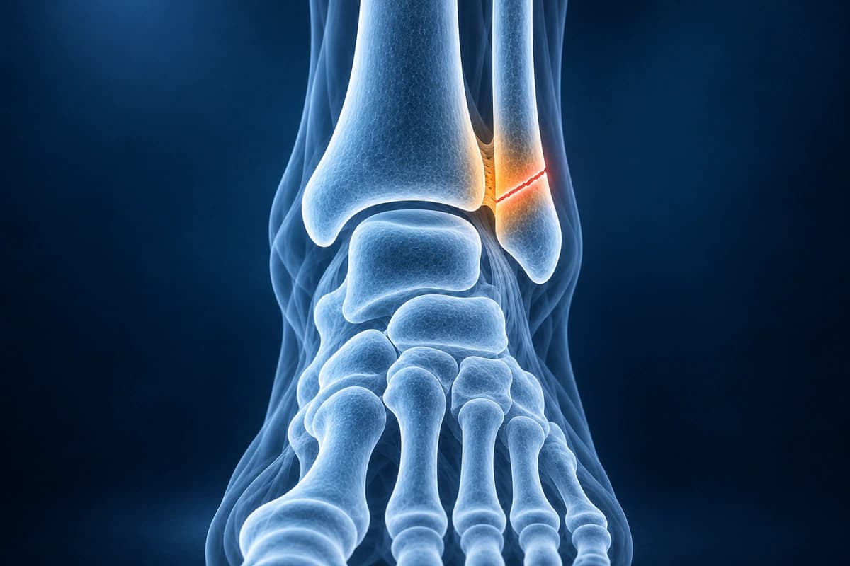
Key Imaging Findings
- Fracture line through the lateral malleolus with Weber classification
- Medial malleolus fracture or deltoid ligament widening
- Posterior malleolus fragment involvement and percentage of articular surface
- Syndesmotic widening suggesting tibiofibular injury
- Talar shift indicating unstable injury pattern
- Occult fractures visible on MRI but not on X-ray
Frequently Asked Questions
When is CT or MRI preferred over plain X-ray for ankle fractures?
Plain radiographs (AP, lateral, mortise views) are the first-line modality for ankle fractures. CT is indicated for comminuted fractures, posterior malleolus assessment, pilon fractures, and pre-operative planning of articular reduction. MRI adds value when X-ray and CT are negative but clinical suspicion remains high — particularly for non-displaced stress fractures, syndesmotic ligament integrity (AITFL, PITFL), and associated osteochondral talar dome injury. STIR sequences are the most sensitive for occult fracture marrow edema.
How does the Lauge-Hansen classification relate to MRI ligament findings?
The Lauge-Hansen system predicts injury sequence based on the deforming force. A supination-adduction pattern injures the lateral ligaments (ATFL, CFL) before the medial malleolus, visible on MRI as lateral ligament disruption with a transverse fibular fracture. Supination-external rotation — the most common pattern — progresses from AITFL disruption through distal fibular spiral fracture to posterior malleolus and then deltoid injury. AI can help explain these classification terms, but it cannot confirm ankle stability. A clinician must review the full imaging study and examination findings.
What MRI signs indicate an unstable ankle mortise requiring surgical fixation?
Medial clear space widening greater than 4 mm on mortise radiographs is the classic surgical threshold, but MRI directly visualises the deep deltoid ligament. Complete deep deltoid disruption on coronal T2-FS — shown as a fluid-filled gap medially — combined with fibular fracture at or above the level of the tibial plafond (Weber B/C) indicates rotational instability requiring operative stabilisation. Intact deep deltoid fibres on MRI may support non-operative management even with equivocal stress-view radiographs.
Sources
- Michelson JD. Ankle fractures resulting from rotational injuries. J Am Acad Orthop Surg. 2003;11(6):403-412.
- Park SS, Kubiak EN, Egol KA, et al. Stress radiographs after ankle fracture: the effect of ankle position and deltoid ligament status on medial clear space measurements. J Orthop Trauma. 2006;20(1):11-18.
- Koval KJ, Egol KA, Cheung Y, et al. Does a positive ankle stress test indicate the need for operative treatment after lateral malleolus fracture? A preliminary report. J Orthop Trauma. 2007;21(7):449-455.
- Expert Panel on Musculoskeletal Imaging; Smith SE, Chang EY, et al. ACR Appropriateness Criteria Acute Trauma to the Ankle. J Am Coll Radiol. 2020;17(11S):S355-S366.
Related Articles
Learn about ATFL and CFL healing potential, conservative versus surgical treatment, and when chronic instability requires intervention.
Your ankle MRI shows an ATFL or CFL sprain or tear? Learn what grades 1, 2 and 3 mean, partial vs complete tears, and how treatment changes by grade.
Ankle MRI cost ranges, what affects the price, insurance coverage, and practical tips to lower your out-of-pocket cost.
Understand your imaging before your next appointment
MRI, X-ray or CT from a CD, folder or ZIP. A PDF report or photos of the films work too.
Free to open and preview, no account needed. An optional AI report from $10 explains your images in plain language, with questions for your doctor.
Your raw files stay on this device. Only rendered images and minimal context are sent when you request an AI report.
Medical Disclaimer: This page is for informational and educational purposes only. It does not constitute medical advice, diagnosis, or treatment. AI-generated analysis may contain errors. Always consult a qualified healthcare professional for medical decisions. Full Disclaimer