
Common Symptoms
- Lateral ankle pain and swelling along the posterior fibula and peroneal groove, distal to the lateral malleolus
- Tenderness on palpation of the peroneus longus or peroneus brevis tendon posterior to the lateral malleolus
- Pain and weakness with resisted ankle eversion, specifically isolating the peroneus brevis insertion at the fifth metatarsal base
- Snapping or subluxation sensation behind the fibula with dorsiflexion and eversion, indicating superior peroneal retinaculum (SPR) laxity
- Swelling within the peroneal tendon sheath evidenced by a fluid-filled ridge posterior to the lateral malleolus
- Chronic lateral instability mimicking recurrent ankle sprains due to dynamic peroneal tendon dysfunction
Peroneal Tendon Tear MRI Signs: Brevis Split, Longus Tear, and Subluxation
Your ankle MRI shows a peroneus brevis split tear, a longus tear or tendon subluxation? Learn what these findings mean and how they link to instability.
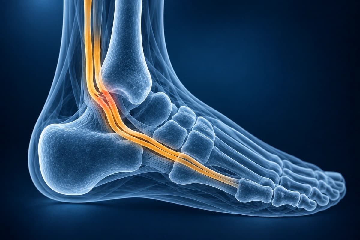
Peroneus Brevis Split Tear: Decoding the Report Wording
A “longitudinal split tear of the peroneus brevis” means one of the two tendons behind the outer ankle bone has split lengthwise, often into a C shape that partly wraps around the peroneus longus. It is a tear along the tendon rather than straight across it. Tenosynovitis and subluxation, often listed nearby in the report, are separate findings.
Upload Ankle ImagingPeroneal phrases you may find in the report
- “Longitudinal split tear of the peroneus brevis tendon”
- “C-shaped” or “bisected peroneus brevis”
- “Intrasubstance signal” or “tendinosis of the peroneal tendons”
- “Peroneal tenosynovitis” or “fluid in the common peroneal sheath”
- “Subluxation of the peroneal tendons” or “stripped superior peroneal retinaculum”
- “Flat” or “convex retromalleolar groove”
Split, tendinosis, tenosynovitis and subluxation compared
A split tear shows on axial images as a cleft through the peroneus brevis, with the two halves forming a C around the peroneus longus. Tendinosis or intrasubstance signal means the tendon is abnormal inside without a clear split. Tenosynovitis means the tendon sheath is inflamed, with fluid around the tendons. Subluxation means the tendons slip out of their groove behind the fibula, usually because the superior peroneal retinaculum that holds them is torn or stripped.
Some appearances can mimic a tear. A normal peroneus brevis can look mildly crescent-shaped or bifurcated, the magic angle effect can brighten a healthy tendon, and an extra peroneus quartus muscle or a low-lying brevis muscle belly can crowd the groove. Radiologists still report these variants because they can predispose to tears.
How much the finding weighs in treatment
Peroneal tendon injuries should be considered in anyone with lasting outer ankle pain, and they are often linked with sprains and ankle instability. Treatment depends on symptoms and activity level more than on the exact wording: people with low physical demands usually do well with conservative care, while those with high demands may benefit from surgery if conservative care fails.
What to ask about the peroneal tendons
- Is the peroneus brevis split along its length, and is the peroneus longus intact?
- Do the tendons stay in their groove, or does the report suggest the retinaculum is torn?
- Could the split explain why my ankle still hurts after the sprain healed?
- Would you try physiotherapy or a brace first, and what would make surgery worth discussing?
Overview
The peroneal tendons (peroneus longus and brevis) run along the lateral ankle and are important for ankle stability and eversion. Injuries include tendon tears (longitudinal splits are most common in peroneus brevis), subluxation or dislocation from the fibular groove, and tendinopathy. These injuries are often associated with lateral ankle sprains and chronic ankle instability. Our AI consortium evaluates tendon morphology, position, and the integrity of the superior peroneal retinaculum.
Peroneal tendon tears are often missed when the search stops at a routine ankle sprain. The key MRI questions are whether the brevis tendon has a longitudinal split, whether the longus tear extends toward the cuboid tunnel, whether the superior peroneal retinaculum is stripped, and whether tendon instability explains persistent lateral ankle pain. Compare the broader ankle MRI reading guide when the report also mentions ligament edema, talar dome cartilage injury, or marrow contusion.
Key Imaging Findings
- Longitudinal split tear of peroneus brevis with C-shaped morphology on axial MRI
- Peroneus longus tear or tendinopathy with increased signal
- Subluxation or dislocation of tendons from the fibular groove
- Superior peroneal retinaculum tear or stripping
- Shallow or convex fibular groove predisposing to subluxation
- Peroneal tendon tenosynovitis with fluid surrounding tendons
Frequently Asked Questions
How does MRI differentiate peroneus brevis split tears from peroneus longus tears?
Axial PD-FS sequences at the level of the retromalleolar groove are the key diagnostic plane. Peroneus brevis split tears present as a C-shaped or bifid tendon with a longitudinal intrasubstance cleft on axial images, often with the peroneus longus wedged within the split. Peroneus longus tears more commonly occur at the cuboid tunnel or os peroneum level — a fragmented or enlarged os peroneum on lateral radiograph and a longitudinal hyperintense cleft on axial PD-FS confirms the diagnosis. Peroneal tendon sheath effusion and SPR signal disruption on coronal images indicate associated retinaculum injury.
What is peroneal tendon subluxation and how is it assessed on imaging?
Peroneal tendon subluxation occurs when the superior peroneal retinaculum (SPR) fails to contain the peroneus brevis and longus within the retromalleolar groove. MRI signs include SPR disruption or periosteal sleeve avulsion from the fibula on axial T2-FS, shallow or convex retromalleolar groove morphology, and tendon displacement anterior to the fibular tip. Dynamic ultrasound with real-time dorsiflexion-eversion stress directly visualises subluxation. AI can help explain the report, but an ankle specialist must confirm tendon instability and decide whether surgery or non-surgical care is appropriate.
When should peroneal tendon pathology be suspected after a lateral ankle sprain?
Peroneal tendon involvement should be suspected when lateral ankle pain persists beyond 6–8 weeks following a sprain, when pain localises to the posterolateral fibula rather than the ATFL footprint, or when eversion strength is disproportionately reduced relative to inversion stability. Studies show that up to 25% of chronic lateral ankle instability cases have concurrent peroneal tendon tears on MRI. AI can help explain terms about the ankle ligaments and peroneal tendons, but it may miss injuries. A clinician must review persistent pain after a sprain.
Sources
- Rosenberg ZS, Beltran J, Cheung YY, et al. MR features of longitudinal tears of the peroneus brevis tendon. AJR Am J Roentgenol. 1997;168(1):141-147.
- Taljanovic MS, Alcala JN, Gimber LH, et al. High-resolution US and MR imaging of peroneal tendon injuries. Radiographics. 2015;35(1):179-199.
- Khoury NJ, el-Khoury GY, Saltzman CL, Kathol MH. Peroneus longus and brevis tendon tears: MR imaging evaluation. Radiology. 1996;200(3):833-841.
- Wang XT, Rosenberg ZS, Mechlin MB, Schweitzer ME. Normal variants and diseases of the peroneal tendons and superior peroneal retinaculum: MR imaging features. Radiographics. 2005;25(3):587-602.
- Eckert WR, Davis EA Jr. Acute rupture of the peroneal retinaculum. J Bone Joint Surg Am. 1976;58(5):670-672.
- van Dijk PA, Miller D, Calder J, et al. The ESSKA-AFAS international consensus statement on peroneal tendon pathologies. Knee Surg Sports Traumatol Arthrosc. 2018;26(10):3096-3107.
Related Articles
Learn when 3T ankle MRI can help for cartilage lesions, peroneal tendons, syndesmosis injuries, and occult fractures, and when X-ray or 1.5T MRI is enough.
Understand your ankle MRI report including ligament evaluation, tendon assessment, and osteochondral lesion detection.
Get an educational explanation of imaging terms related to Peroneal Tendon Tear
Upload your MRI or X-ray DICOM files for private, AI-powered analysis. 4 models analyze independently — raw scan files stay in your browser.
Upload & AnalyzeMedical Disclaimer: This page is for informational and educational purposes only. It does not constitute medical advice, diagnosis, or treatment. AI-generated analysis may contain errors. Always consult a qualified healthcare professional for medical decisions. Full Disclaimer