Posterior Tibial Tendon Dysfunction on MRI & X-ray: Imaging Findings
Posterior tibial tendon dysfunction (PTTD) is the most common cause of acquired adult flatfoot deformity. The posterior tibial tendon supports the medial longitudinal arch and controls hindfoot inversion during gait. When the tendon degenerates, it progressively elongates and loses its mechanical advantage, leading to arch collapse and hindfoot valgus. PTTD is staged from I to IV: Stage I involves tendinopathy with a normal arch, Stage II shows flexible flatfoot deformity, Stage III presents rigid deformity with subtalar arthritis, and Stage IV adds ankle valgus tilt. MRI is essential for evaluating tendon integrity, staging the disease, and assessing associated spring ligament damage.
Common Symptoms
- Medial ankle and midfoot pain along the course of the posterior tibial tendon (PTT) from the medial malleolus to the navicular
- Progressive flatfoot deformity — loss of the medial longitudinal arch visible on standing assessment
- Inability to perform a single-leg heel rise, or pain and asymmetric heel valgus during the attempt
- Swelling along the medial ankle behind the medial malleolus within the PTT sheath
- Too-many-toes sign — excess lateral toe visibility when the patient is viewed from behind due to hindfoot valgus and forefoot abduction
- Late-stage rigid flatfoot with subtalar and midtarsal joint arthritis causing hindfoot pain at rest
What We Look For on Imaging
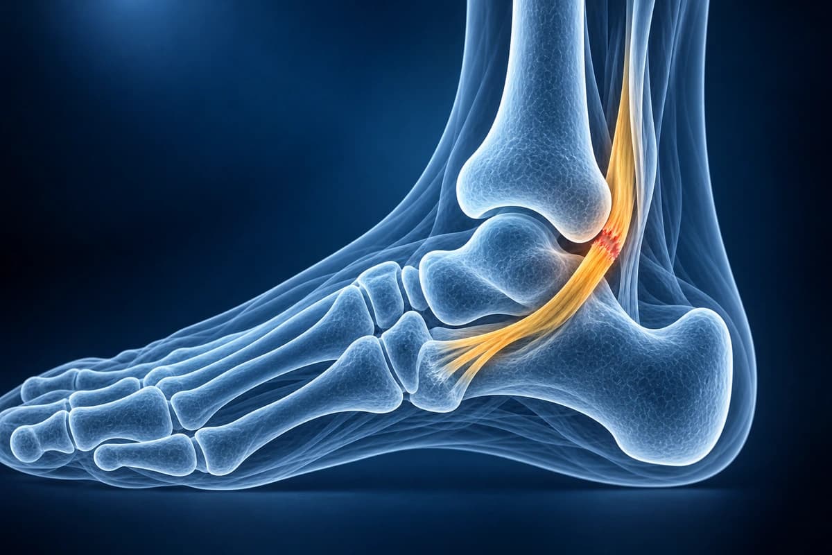
- Tendon thickening or thinning with increased intratendinous signal on MRI
- Longitudinal split tears of the posterior tibial tendon on axial images
- Spring ligament (superomedial calcaneonavicular) attenuation or tear
- Peritendinous tenosynovitis with fluid surrounding the tendon sheath
- Hindfoot valgus alignment and talar head uncovering on weight-bearing views
- Sinus tarsi edema and subtalar joint degenerative changes in advanced stages
Frequently Asked Questions
What MRI sequences and findings confirm posterior tibial tendon dysfunction (PTTD)?
MRI can show posterior tibial tendon tears, longitudinal splits, tendon degeneration, and surrounding fluid or edema.
How does AI-assisted MRI staging of PTTD guide surgical planning?
Treatment planning depends on tendon damage, arch flexibility, alignment and arthritis, assessed through examination and imaging. AI cannot assign a clinical stage or choose surgery.
Can PTTD be distinguished from medial ankle tendinopathy of other tendons on MRI?
Yes. The medial ankle contains the Tom, Dick, and Harry tendons — tibialis posterior, FDL, and flexor hallucis longus (FHL) — in the tarsal tunnel. Axial sequences clearly separate each tendon by position: PTT is the most anterior and largest, directly behind the medial malleolus, inserting on the navicular tuberosity. FHL lies posteriorly between the medial and lateral talar processes — isolated FHL tenosynovitis produces posterior ankle pain and hallux triggering. AI can help explain which tendon a report refers to, but a clinician must confirm the findings and decide whether an injection or surgery is appropriate.
Sources
- Johnson KA, Strom DE. Tibialis posterior tendon dysfunction. Clin Orthop Relat Res. 1989;(239):196-206.
- Rosenberg ZS, Cheung Y, Jahss MH, et al. Rupture of posterior tibial tendon: CT and MR imaging with surgical correlation. Radiology. 1988;169(1):229-235.
- Balen PF, Helms CA. Association of posterior tibial tendon injury with spring ligament injury, sinus tarsi abnormality, and plantar fasciitis on MR imaging. AJR Am J Roentgenol. 2001;176(5):1137-1143.
Understand your imaging before your next appointment
MRI, X-ray or CT from a CD, folder or ZIP. A PDF report or photos of the films work too.
Free to open and preview, no account needed. An optional AI report from $10 explains your images in plain language, with questions for your doctor.
Your raw files stay on this device. Only rendered images and minimal context are sent when you request an AI report.
Medical Disclaimer: This page is for informational and educational purposes only. It does not constitute medical advice, diagnosis, or treatment. AI-generated analysis may contain errors. Always consult a qualified healthcare professional for medical decisions. Full Disclaimer