Achilles Tendon Tear on MRI: Imaging Findings
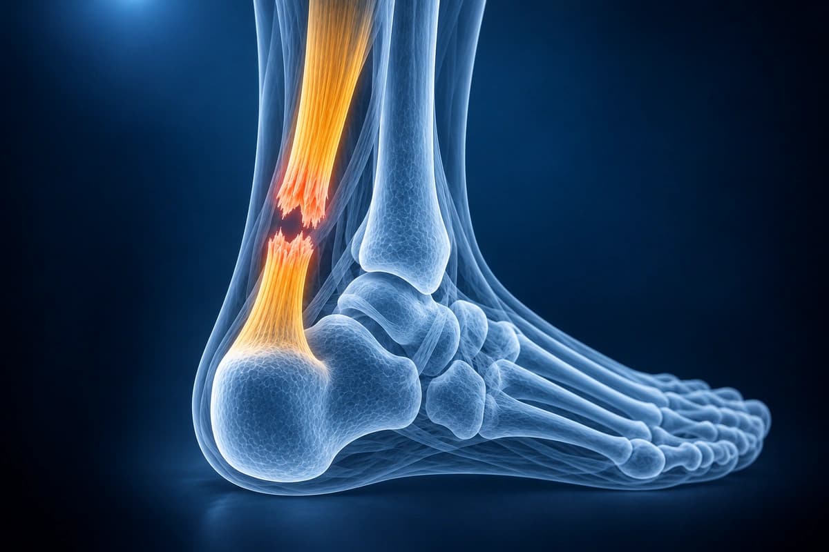
The Achilles tendon is the strongest and thickest tendon in the body, but is also one of the most commonly ruptured tendons. Tears typically occur 2-6 cm above the calcaneal insertion in the watershed zone of reduced blood supply. MRI provides excellent visualization of tear location, extent, gap size, and tendon quality.
Common Symptoms
- Sudden sharp pain at the posterior heel or mid-substance Achilles tendon, often described as a "pop"
- Palpable gap in the Achilles tendon approximately 2–6 cm proximal to the calcaneal insertion
- Positive Thompson squeeze test — absence of plantarflexion when the calf is compressed
- Significant swelling and ecchymosis along the posterior ankle and heel
- Inability to perform a single-leg heel rise or push off during gait
- Weakness in plantarflexion with preserved passive range of motion in acute complete rupture
Key Imaging Findings
- Complete disruption of tendon fibers with visible gap on sagittal MRI
- High T2 signal within the tendon substance indicating edema or tear
- Tendon thickening with heterogeneous signal in chronic tendinopathy
- Retraction of torn tendon ends with measurement of gap distance
- Partial tear with intact fibers maintaining continuity
- Surrounding paratenon edema and hemorrhage
Frequently Asked Questions
What MRI findings differentiate partial from complete Achilles tendon rupture?
On sagittal T2-fat-saturated or STIR sequences, partial tears show focal intratendinous hyperintensity with maintained tendon continuity and increased AP diameter, while complete ruptures display a fluid-filled gap between proximal and distal stumps. The tendon gap size, measured in millimetres on sagittal images, guides surgical planning. Axial PD-FS images clarify the cross-sectional involvement percentage, distinguishing central-core versus peripheral partial tears. Peritendinous fluid and reactive edema in Kager's fat pad are consistent secondary findings.
Can ultrasound replace MRI for diagnosing Achilles tendon tears?
Dynamic ultrasound with real-time Thompson maneuver is highly accurate (>90% sensitivity) for complete ruptures and is lower cost and immediately available. However, MRI surpasses ultrasound in characterising partial tears, quantifying tendon substance involvement, assessing insertional enthesopathy, and evaluating concurrent retrocalcaneal bursitis or paratenon inflammation. MRI is preferred pre-operatively and in cases where ultrasound findings are equivocal, particularly for mid-substance versus insertional tear distinction critical to surgical approach.
How does AI analysis of Achilles MRI assist in return-to-sport decisions?
An Achilles MRI may show changes in tendon thickness, structure, and signal during recovery. AI can help explain the wording in your report, but it cannot measure tendon strength, confirm that healing is complete, or decide when returning to sport is safe. Your treating clinician must guide that decision.
Sources
- Carr AJ, Norris SH. The blood supply of the calcaneal tendon. J Bone Joint Surg Br. 1989;71(1):100-101.
- Maffulli N. The clinical diagnosis of subcutaneous tear of the Achilles tendon. A prospective study in 174 patients. Am J Sports Med. 1998;26(2):266-270.
- Hartgerink P, Fessell DP, Jacobson JA, van Holsbeeck MT. Full- versus partial-thickness Achilles tendon tears: sonographic accuracy and characterization in 26 cases with surgical correlation. Radiology. 2001;220(2):406-412.
Related Articles
Complete ankle sprain rehabilitation protocol including RICE, proprioception training, strengthening, and return-to-sport criteria.
Understand common ankle conditions including ligament sprains, fractures, Achilles tendon tears, peroneal injuries, and osteochondral defects.
Ankle MRI cost ranges, what affects the price, insurance coverage, and practical tips to lower your out-of-pocket cost.
Understand your imaging before your next appointment
MRI, X-ray or CT from a CD, folder or ZIP. A PDF report or photos of the films work too.
Free to open and preview, no account needed. An optional AI report from $10 explains your images in plain language, with questions for your doctor.
Your raw files stay on this device. Only rendered images and minimal context are sent when you request an AI report.
Medical Disclaimer: This page is for informational and educational purposes only. It does not constitute medical advice, diagnosis, or treatment. AI-generated analysis may contain errors. Always consult a qualified healthcare professional for medical decisions. Full Disclaimer