
Tarsal Tunnel Syndrome on MRI & X-ray: Imaging Findings
Tarsal tunnel syndrome is a compressive neuropathy of the tibial nerve or its branches as they pass through the tarsal tunnel beneath the flexor retinaculum on the medial ankle. The tarsal tunnel contains the tibial nerve, posterior tibial artery and veins, and the tendons of tibialis posterior, flexor digitorum longus, and flexor hallucis longus. Compression can result from space-occupying lesions such as ganglion cysts, varicosities, accessory muscles, or lipomas, as well as from post-traumatic fibrosis, tenosynovitis, or hindfoot valgus deformity. MRI is valuable for identifying structural causes of nerve compression and evaluating nerve morphology.
Common Symptoms
- Burning, tingling, or electric pain radiating from the medial ankle along the plantar surface of the foot and toes
- Positive Tinel sign — percussion over the tarsal tunnel posterior to the medial malleolus reproduces distal paresthesias
- Nocturnal worsening of plantar foot pain and numbness, partially relieved by dependent position
- Symptoms reproduced by prolonged standing or walking, with relief on rest and elevation
- Intrinsic foot muscle weakness in chronic cases — interosseous atrophy causing subtle toe spread deficit
- Medial heel numbness from the calcaneal branch of the posterior tibial nerve, or lateral plantar nerve distribution deficits
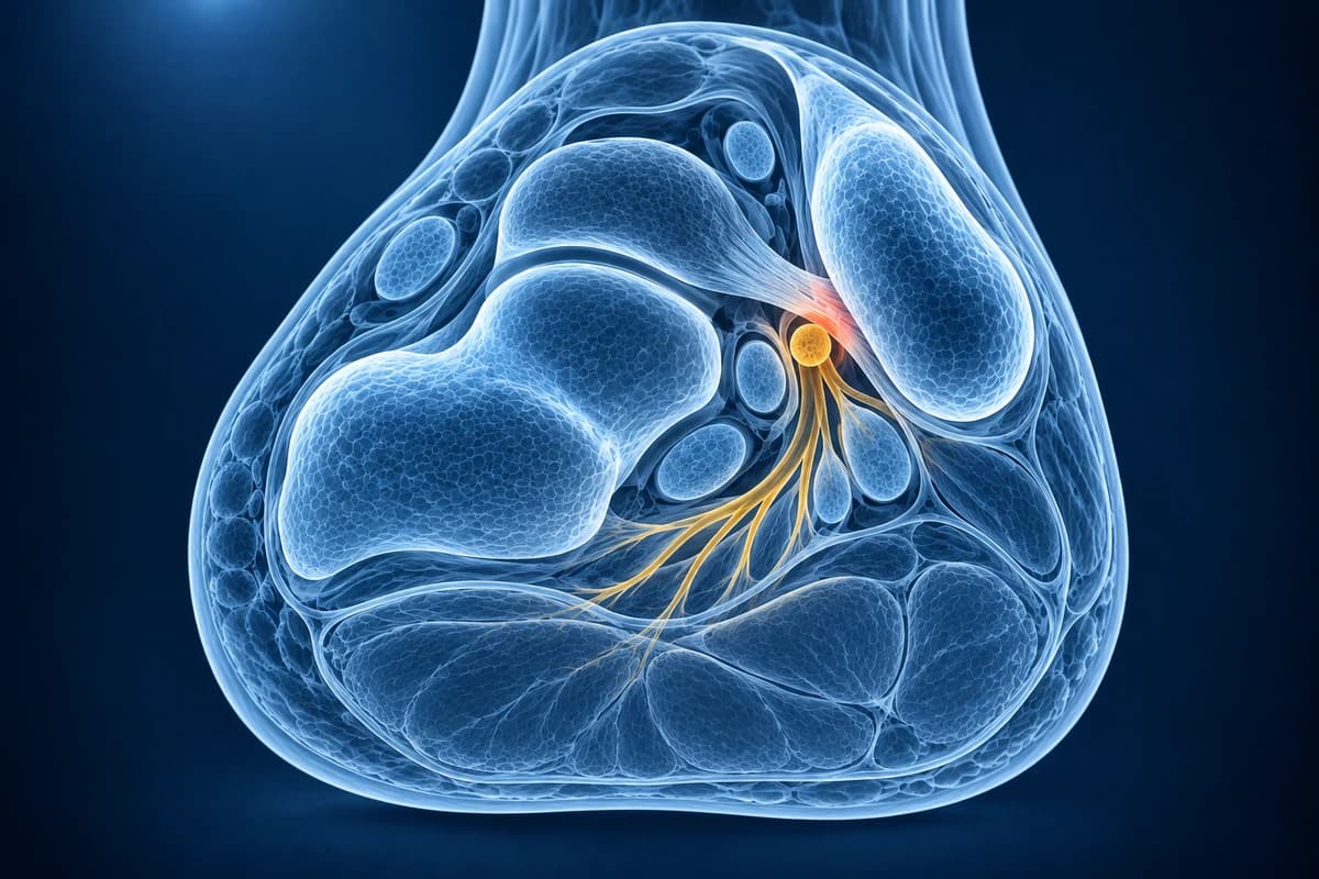
What We Look For on Imaging
- Space-occupying lesions within the tarsal tunnel such as ganglion cysts or lipomas
- Varicose veins or venous malformations compressing the tibial nerve
- Tibial nerve enlargement or increased T2 signal indicating neuropathy
- Flexor tenosynovitis narrowing the available space within the tunnel
- Accessory muscles (such as accessory flexor digitorum longus) occupying the tunnel
- Denervation edema in the plantar intrinsic muscles on fluid-sensitive sequences
Frequently Asked Questions
What does MRI reveal in tarsal tunnel syndrome and when is it essential?
MRI is essential when clinical examination and nerve conduction studies are inconclusive or when a space-occupying lesion is suspected. Axial T1 and T2-FS sequences through the tarsal tunnel evaluate the posterior tibial nerve, medial and lateral plantar nerve divisions, and the contents of the flexor retinaculum tunnel. Common MRI findings include ganglion cyst (high T2, well-defined), lipoma (T1 hyperintense), varicose veins (serpiginous T2-bright structures), or accessory muscles (isointense to muscle on all sequences) compressing the nerve. Neural signal change — T2 hyperintensity within the posterior tibial nerve — indicates intrinsic nerve injury.
How can AI analysis of ankle MRI improve tarsal tunnel syndrome diagnosis?
MRI can help a clinician look for changes in the tibial nerve and nearby structures at the inner ankle. AI can help explain report wording, but it cannot confirm nerve compression or identify the cause of your symptoms. A clinician must interpret the scan alongside an examination and any other tests.
What is the role of imaging in differentiating tarsal tunnel syndrome from plantar fasciitis?
Plantar fasciitis manifests as plantar heel pain with maximum tenderness at the calcaneal insertion of the fascia, typically worst with first steps in the morning. MRI shows plantar fascia thickening (>4 mm), perifascicular edema, and calcaneal enthesopathy on sagittal STIR. Tarsal tunnel syndrome produces burning medial ankle and plantar paresthesias with a positive Tinel sign and no morning stiffness pattern. These findings can coexist. AI can help explain their names, but it cannot establish which finding causes your symptoms or determine which treatment will help.
Sources
- Ahmad M, Tsang K, Mackenney PJ, Adedapo AO. Tarsal tunnel syndrome: a literature review. Foot Ankle Surg. 2012;18(3):149-152.
- Erickson SJ, Quinn SF, Kneeland JB, et al. MR imaging of the tarsal tunnel and related spaces: normal and abnormal findings with anatomic correlation. AJR Am J Roentgenol. 1990;155(2):323-328.
- Frey C, Kerr R. Magnetic resonance imaging and the evaluation of tarsal tunnel syndrome. Foot Ankle. 1993;14(3):159-164.
- Berkowitz JF, Kier R, Rudicel S. Plantar fasciitis: MR imaging. Radiology. 1991;179(3):665-667.
Understand your imaging before your next appointment
MRI, X-ray or CT from a CD, folder or ZIP. A PDF report or photos of the films work too.
Free to open and preview, no account needed. An optional AI report from $10 explains your images in plain language, with questions for your doctor.
Your raw files stay on this device. Only rendered images and minimal context are sent when you request an AI report.
Medical Disclaimer: This page is for informational and educational purposes only. It does not constitute medical advice, diagnosis, or treatment. AI-generated analysis may contain errors. Always consult a qualified healthcare professional for medical decisions. Full Disclaimer