Lateral Ankle Ligament Grading: ATFL & CFL Tears on MRI
Grade 1, 2, and 3 lateral ankle ligament injuries — ATFL and CFL anatomy, MRI signs of partial vs complete tears, and treatment by grade.
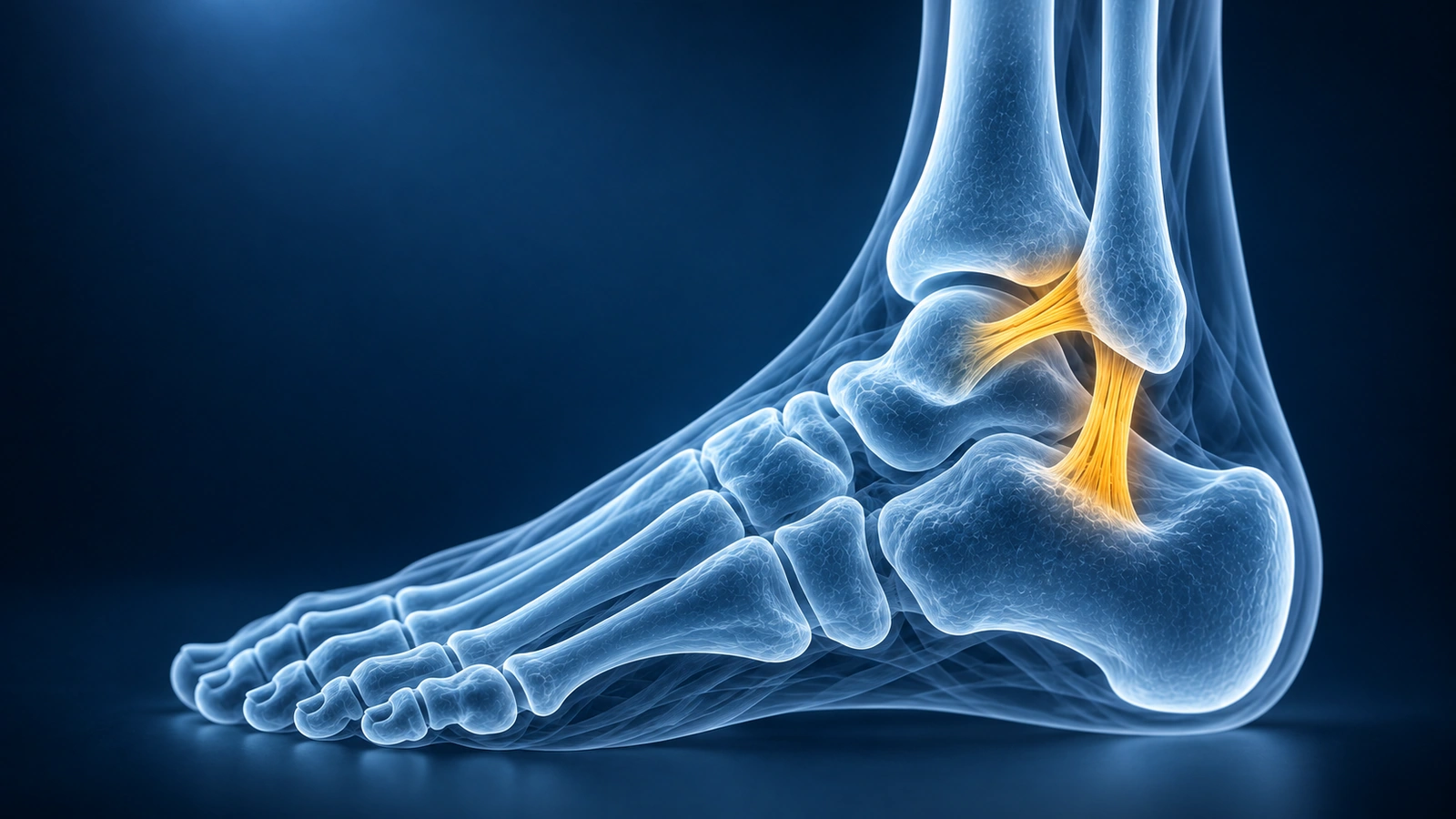
The lateral ankle ligament complex is the most commonly injured structure in the body, with millions of ankle sprains occurring each year. Three ligaments form this complex — the anterior talofibular ligament (ATFL), the calcaneofibular ligament (CFL), and the posterior talofibular ligament (PTFL). The ATFL runs from the anterior border of the fibula to the lateral aspect of the talar neck and is the weakest and most frequently torn of the three. The CFL extends from the fibular tip to the lateral surface of the calcaneus and is the second most commonly injured ligament. The PTFL is rarely torn in isolation, typically only in severe dislocations.
When an ankle sprain is evaluated on MRI, injuries are graded 1 through 3 based on the degree of fiber disruption. This grading directly guides treatment decisions, determines return-to-sport timelines, and identifies patients at risk for chronic ankle instability if the injury is undertreated. Understanding what each grade means on MRI helps you interpret your findings and have an informed conversation with your orthopedic surgeon or sports medicine physician.
Grade 1: Ligament Sprain
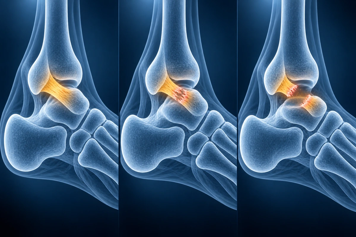
A Grade 1 lateral ankle injury is a mild sprain in which the ATFL or CFL fibers are stretched but structurally intact. No macroscopic fiber tearing has occurred. On MRI, the ligament shows increased signal on fluid-sensitive sequences (T2 or STIR), reflecting edema and periligamentous soft-tissue swelling, but the fiber continuity is preserved from origin to insertion without any visible gap or discontinuity. The ligament may appear mildly thickened or surrounded by edema, but the internal architecture remains organized.
Clinically, the anterior drawer test is negative or shows minimal laxity compared to the contralateral ankle. Patients experience localized lateral tenderness and swelling but no mechanical giving way. Recovery with conservative management — rest, ice, compression, and elevation followed by proprioceptive rehabilitation — typically takes 1 to 3 weeks. Return to sport is expected without residual instability.
Grade 2: Partial Tear
A Grade 2 injury involves a partial tear of the ligament — some fibers are disrupted while others remain intact. On MRI, the ligament demonstrates irregular contour, thinning, or focal fiber discontinuity, but a continuous portion can still be traced along part of the ligament's course. T2 signal is markedly elevated within the substance of the ligament, and surrounding soft-tissue edema is more prominent than in a Grade 1 injury. The ligament may appear lax or wavy rather than the normal taut, low-signal band.
Clinically, the anterior drawer test shows increased laxity with a soft or delayed endpoint, reflecting partial loss of mechanical restraint. Patients often report a sensation of the ankle giving way on uneven surfaces. Recovery with structured rehabilitation typically takes 4 to 8 weeks, though athletes in high-demand sports may require longer. Persistent instability after Grade 2 injuries that do not respond to conservative care may warrant consideration of surgical repair or augmentation.
Grade 3: Complete Tear
A Grade 3 injury is a complete tear with full disruption of the ligament fibers. On MRI, this appears as frank discontinuity of the ligament — the normal dark, taut band is absent and replaced by a gap filled with fluid signal. The ligament may appear as a wavy, retracted, or amorphous low-signal structure with surrounding hemorrhage and edema. In some cases the ligament is virtually invisible on standard sequences, a pattern sometimes described as an empty or absent fiber pattern. For guidance on interpreting these MRI findings, our ankle MRI reading guide walks through the key sequences and landmarks.
Clinically, the anterior drawer test shows gross laxity with no firm endpoint. The talar tilt test is also positive when the CFL is involved. Recovery from a Grade 3 ATFL tear in a sedentary or low-demand patient can be managed conservatively with a functional brace or cast for 4 to 6 weeks followed by rehabilitation, with full recovery in 8 to 12 weeks. However, athletes in pivoting and cutting sports who experience persistent instability after appropriate rehabilitation are candidates for surgical repair (Broström procedure) or ligament reconstruction.
ATFL vs CFL Injury Patterns
The mechanism of lateral ankle sprains follows a predictable sequential pattern. The ATFL is the primary restraint to anterior talar translation when the ankle is in plantarflexion — the position it occupies at the moment of a typical inversion sprain. As a result, the ATFL is the first ligament to fail and is injured in isolation in most mild-to-moderate sprains. With greater inversion force, the CFL — which resists both inversion talar tilt and anterior translation in the neutral position — is torn next, producing a combined ATFL and CFL injury that significantly increases ankle instability.
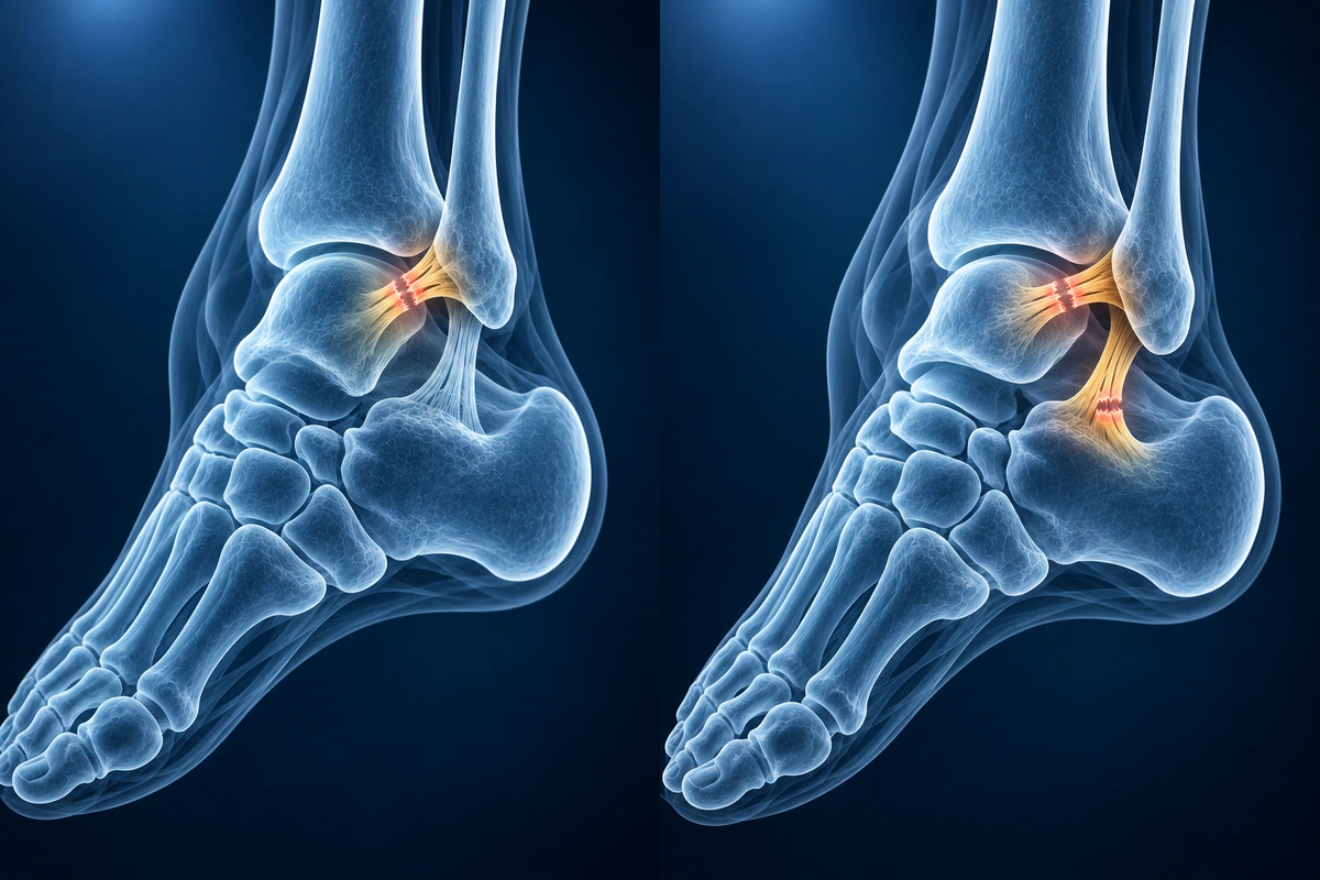
Combined ATFL and CFL tears are associated with greater mechanical laxity, higher rates of chronic instability, and a greater likelihood of associated injuries. On MRI, always evaluate the peroneal tendons alongside the lateral ligaments — longitudinal peroneal tendon tears, particularly of the peroneus brevis, occur in up to 25% of significant lateral ankle sprains. In high-energy mechanisms, also assess the syndesmosis (the anterior and posterior inferior tibiofibular ligaments) for a high ankle sprain component, which requires longer immobilization and may need surgical stabilization. If undertreated, combined lateral ligament injuries frequently lead to chronic lateral ankle instability, characterized by recurrent giving way, proprioceptive deficits, and accelerated cartilage wear. To understand whether surgery is necessary, our guide on ankle ligament healing without surgery covers the evidence on conservative versus surgical outcomes.
When MRI Is Indicated
The Ottawa Ankle Rules identify when X-ray is required after an ankle injury to rule out fracture — bony tenderness at the posterior edge of the fibula or tibia, or inability to weight bear. MRI is not the first-line investigation for every ankle sprain. It is indicated when pain and functional limitation persist beyond 4 to 6 weeks despite appropriate conservative management, when clinical examination suggests a high-grade injury with significant instability, or when an associated injury is suspected — such as a peroneal tendon tear, osteochondral lesion of the talus, or syndesmotic injury.
In athletes or patients considering surgical repair, MRI is used preoperatively to confirm ligament grade, characterize the tear morphology (mid-substance vs avulsion), identify any associated peroneal tendon pathology, and assess the articular cartilage of the tibiotalar joint. MRI findings directly influence the surgical approach — an avulsion tear at the fibular attachment may be repaired directly, while a mid-substance tear with attenuated tissue may require augmentation with allograft or tendon transfer.
Key Takeaways
- The ATFL runs from the anterior fibula to the talar neck and is the first ligament injured in a lateral ankle sprain; the CFL runs from the fibular tip to the calcaneus and fails with greater inversion force
- Grade 1 (sprain): edema around intact fibers on MRI, no discontinuity, recovery 1–3 weeks with conservative treatment
- Grade 2 (partial tear): irregular contour and focal fiber disruption on MRI, soft endpoint on drawer test, recovery 4–8 weeks
- Grade 3 (complete tear): frank ligament discontinuity or absent fiber pattern on MRI, gross laxity, recovery 8–12+ weeks; athletes may need the Broström repair
- Combined ATFL and CFL tears greatly increase instability risk; always evaluate peroneal tendons and syndesmosis on the same MRI
- Undertreated Grade 2–3 injuries frequently progress to chronic lateral ankle instability with recurrent giving way and cartilage damage
Frequently Asked Questions
Can I tell the grade of my ankle sprain from symptoms alone?
Symptom severity correlates roughly with grade but is not reliable for grading individual injuries. Swelling, bruising, and pain can be pronounced even in Grade 1 sprains, while some Grade 3 tears cause surprisingly little pain initially because the nerve endings in the ligament are also disrupted. Clinical examination with the anterior drawer and talar tilt tests, combined with MRI when indicated, is required to determine the true grade.
What does a complete ATFL tear look like on MRI?
On fluid-sensitive sequences (T2 or STIR), a complete ATFL tear appears as a gap or zone of bright fluid signal where the ligament should be. The normal dark, taut band from the anterior fibula to the talar neck is absent or replaced by a wavy, retracted, or amorphous structure. Surrounding soft-tissue edema and joint effusion are typically present. In chronic tears the ligament may appear thickened, irregular, or replaced by scar tissue rather than showing an acute fluid gap.
Do Grade 3 ankle ligament tears always require surgery?
No. The majority of Grade 3 ATFL tears — even in recreational athletes — heal sufficiently with functional rehabilitation to allow a return to normal activity without chronic instability. Surgery is reserved for patients who continue to experience recurrent giving way or functional instability after at least 3 to 6 months of structured rehabilitation including proprioceptive training. Competitive athletes in high-demand pivoting sports may be offered earlier surgical repair to minimize time away from sport.
What is chronic lateral ankle instability?
Chronic lateral ankle instability is defined as recurrent episodes of the ankle giving way on uneven terrain, during sport, or with daily activities, persisting for more than 12 months after the initial sprain. It results from inadequate healing of the ATFL or combined ATFL and CFL, leading to persistent mechanical laxity and impaired proprioceptive feedback from the lateral ligament complex. Left untreated, chronic instability accelerates cartilage wear in the tibiotalar joint and increases the risk of osteochondral lesions of the talus.
How is a peroneal tendon tear related to a lateral ankle sprain?
The peroneal tendons — particularly the peroneus brevis — run posterior to the fibula in close proximity to the lateral ligaments. During a forceful inversion sprain, the peroneal tendons contract reflexively to resist the inversion force, and this sudden eccentric load can cause a longitudinal split tear of the peroneus brevis at the level of the fibular groove. Because the symptoms of a peroneal tear (lateral ankle pain, weakness with eversion) overlap with those of a ligament sprain, it is commonly missed on initial examination. MRI reliably distinguishes between the two and should be requested when lateral ankle pain persists despite an apparently straightforward sprain.
Related Articles
Understand common ankle conditions including ligament sprains, fractures, Achilles tendon tears, peroneal injuries, and osteochondral defects.
Learn about ATFL and CFL healing potential, conservative versus surgical treatment, and when chronic instability requires intervention.
Understand your ankle MRI report including ligament evaluation, tendon assessment, and osteochondral lesion detection.
Related Conditions
Ready to analyze your imaging? Upload your MRI or X-ray for AI-powered analysis
Upload your MRI or X-ray DICOM files for private, AI-powered analysis. 4 models analyze independently — raw scan files stay in your browser.
Start AnalysisMedical Disclaimer: This page is for informational and educational purposes only. It does not constitute medical advice, diagnosis, or treatment. AI-generated analysis may contain errors. Always consult a qualified healthcare professional for medical decisions. Full Disclaimer