Common Symptoms
- Pain above the ankle joint, proximal to the lateral malleolus
- Positive squeeze test — pain reproduced by compressing the tibia and fibula together at mid-calf level
- Pain with external rotation stress test of the foot with the knee flexed at 90 degrees
- Slower recovery than a typical lateral ankle sprain — ongoing pain and instability beyond 6 weeks
- Difficulty and pain with push-off during walking or running
- Swelling above the malleoli rather than around the lateral ankle
High Ankle Sprain on MRI: AI-Powered Imaging Analysis
AI-powered high ankle sprain (syndesmosis) analysis on MRI. Understand AITFL and PITFL injuries, interosseous membrane disruption, and how similar findings differ from lateral ankle sprain.
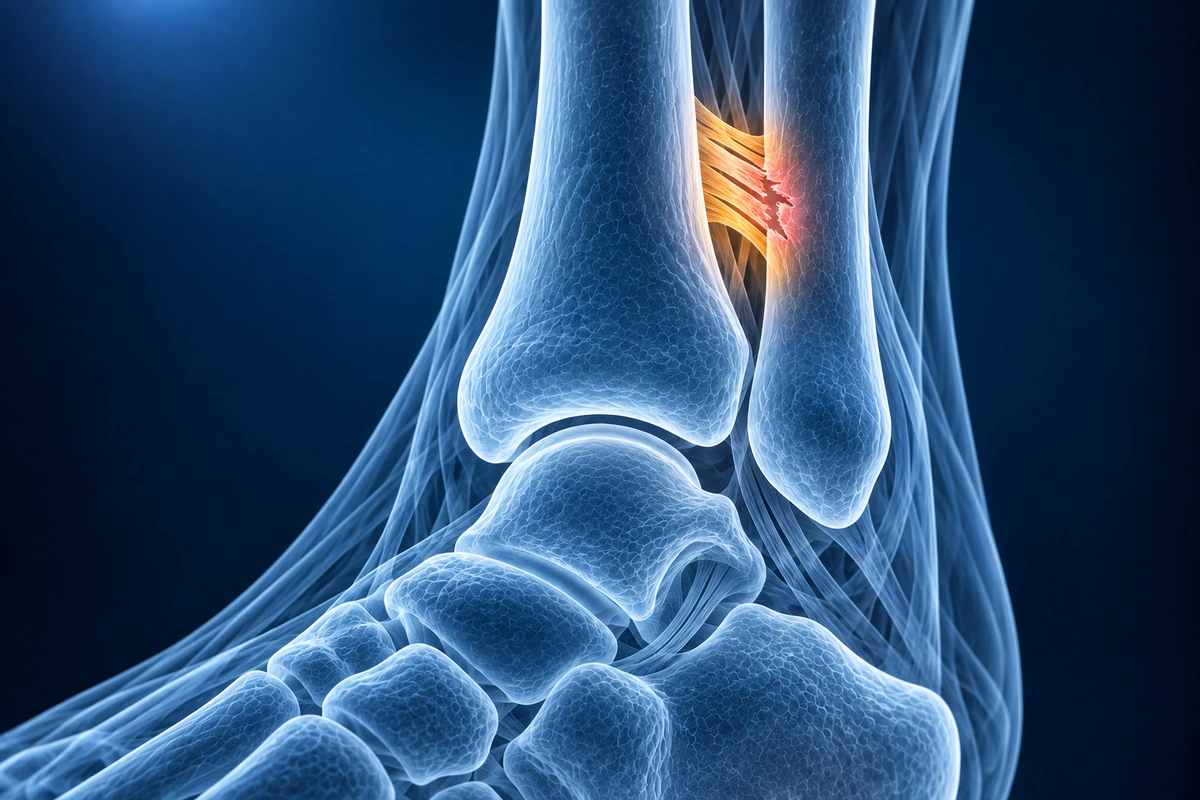
A high ankle sprain is an injury to the syndesmotic ligament complex that connects the tibia and fibula above the ankle joint. The key structures involved are the anterior inferior tibiofibular ligament (AITFL), the posterior inferior tibiofibular ligament (PITFL), and the interosseous membrane — a dense fibrous sheet running between the two bones along their full length. Unlike a common lateral ankle sprain, which tears the ATFL below the ankle joint, a high ankle sprain disrupts the tibiofibular mortise that holds the ankle stable during weight-bearing.
The typical mechanism is external rotation of the foot relative to the leg combined with dorsiflexion — the exact forces in a football tackle where a player's foot is planted and the body rotates over it, or in a ski crash where the boot locks the foot while the leg twists. High ankle sprains account for roughly 1–11% of all ankle sprains in the general population but are significantly more common in contact athletes. Because clinicians sometimes mistake them for severe lateral sprains, they are under-diagnosed and can lead to chronic instability or post-traumatic arthritis if untreated.
Recovery is substantially longer than for a typical lateral sprain. Grade I injuries (AITFL sprain without frank diastasis) typically require 4–8 weeks of protected weight-bearing followed by sport-specific rehabilitation. Grade II injuries (partial AITFL tear with early tibiofibular widening) may take 8–12 weeks. Grade III injuries with complete ligament disruption, significant tibiofibular diastasis, or an associated Maisonneuve fracture — a spiral fracture of the proximal fibula from the same rotational force — frequently require surgical stabilization with a syndesmotic screw or suture button device, followed by a recovery of 3–6 months.
What We Look For on Imaging
- AITFL discontinuity or T2 signal hyperintensity on axial MRI at the anterior aspect of the tibiofibular joint
- Widened tibiofibular clear space on axial images — greater than 6 mm between the medial fibula and the incisura fibularis of the tibia
- Interosseous membrane edema appearing as feathery high T2 signal between the tibia and fibula on coronal sequences
- Posterior malleolus bone contusion or fracture fragment from PITFL avulsion
- Deltoid ligament edema or deep deltoid disruption in severe syndesmotic injuries with medial instability
- Fibular displacement on axial reformats — external rotation or posterior translation of the fibula within the incisura confirms mortise instability
Frequently Asked Questions
How does a high ankle sprain look different from a low ankle sprain on MRI?
A low (lateral) ankle sprain involves the ATFL and CFL below the level of the ankle joint. On axial MRI, the ATFL is injured and T2 signal abnormality is concentrated at the anterior talofibular region. A high ankle sprain shows T2 signal abnormality at the anterior tibiofibular joint — the AITFL — which is proximal and posterior to the ATFL on axial sequences. Additional findings of interosseous membrane edema on coronal images and tibiofibular clear space widening clinch the diagnosis. Both injuries can coexist: a combined lateral and syndesmotic injury has a worse prognosis than either alone.
Do I need surgery for a high ankle sprain?
Whether surgery is needed depends on mortise stability. Stable injuries — where the tibiofibular joint does not widen under stress X-ray — are typically managed non-operatively with a short-leg cast or a controlled ankle motion (CAM) boot and protected weight-bearing. Unstable injuries with frank tibiofibular diastasis, or those associated with a Maisonneuve fracture (a spiral proximal fibula fracture), require surgical stabilization to restore the mortise and prevent chronic instability and post-traumatic arthritis. Stress radiographs under anesthesia are sometimes needed to confirm instability when MRI findings are borderline.
How long does recovery from a high ankle sprain take?
Recovery is significantly longer than for a lateral ankle sprain, which many patients find surprising. A stable Grade I injury with AITFL sprain but no diastasis typically takes 6–8 weeks before return to sport. Grade II injuries with partial ligament disruption often require 10–12 weeks. Grade III injuries managed surgically — with a syndesmotic screw or suture button device — have a recovery arc of 3 to 6 months, including screw removal (if a metal screw was used) before full weight-bearing rehabilitation can begin. Athletes who return too early risk recurrent instability and cartilage injury.
Upload your imaging to check for High Ankle Sprain
Upload your MRI or X-ray DICOM files for private, AI-powered analysis. 4 models analyze independently — raw scan files stay in your browser.
Upload & AnalyzeMedical Disclaimer: This page is for informational and educational purposes only. It does not constitute medical advice, diagnosis, or treatment. AI-generated analysis may contain errors. Always consult a qualified healthcare professional for medical decisions. Full Disclaimer