Osteochondral Lesion of the Talus MRI Signs and Talar Dome Staging
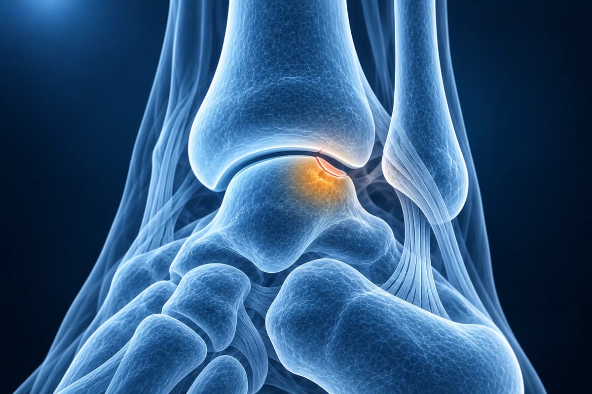
“Bone marrow edema of the talar dome” means the bone under the cartilage of the talus shows fluid-like signal on MRI. On its own, it can be a bone bruise or the earliest stage of an osteochondral lesion. A cartilage defect, subchondral cyst or loose fragment alongside it points to an osteochondral lesion, staged by how stable it is.
Common Symptoms
- Deep, poorly localised ankle pain exacerbated by weight-bearing and relieved by rest
- Joint-line tenderness reproducible on palpation of the anteromedial or anterolateral talar dome with the ankle in plantarflexion
- Intermittent ankle swelling without a clear traumatic event, suggesting chronic intra-articular irritation
- Mechanical symptoms including clicking, catching, or locking if a loose osteochondral fragment is present
- Stiffness and reduced range of motion, particularly in dorsiflexion, after periods of inactivity
- Persistent ankle symptoms beyond 6 weeks following ankle sprain that fail to resolve with standard rehabilitation
Talar Dome Bone Marrow Edema and Osteochondral Lesion in Your Report
Talar dome wording in ankle MRI reports
- “Bone marrow edema” or “edema-like signal in the medial talar dome”
- “Osteochondral lesion of the talus (OLT)” or “osteochondritis dissecans of the talar dome”
- “Focal chondral defect measuring 8 x 6 mm”
- “Subchondral cyst beneath the lesion”
- “Fluid undermining the fragment” or “unstable-appearing fragment”
- “Loose body in the anterior ankle joint”
How the lesion is staged on MRI
Hepple's MRI classification is widely used. Stage 1 is cartilage damage only. Stage 2a is cartilage injury with an underlying fracture and surrounding bone edema, and 2b the same without edema. Stage 3 is a detached but undisplaced fragment, stage 4 a displaced fragment, and stage 5 a subchondral cyst. Older reports may use the Anderson or Berndt and Harty stages instead.
Edema is not the same as damaged cartilage, and edema alone does not mean a fragment is loose. What radiologists look for is fluid signal at the junction between the fragment and the talus: a partly attached fragment shows an irregular bright zone there, and a detached one is surrounded by a complete ring of fluid.
Why stability and size shape treatment
Treatment depends mainly on whether the fragment is attached, how large the lesion is and whether the cartilage is damaged. Non-operative care works best in selected children and teenagers, while adults more often need surgery. In one study of arthroscopic marrow stimulation, lesions of about 150 mm² or more on MRI failed far more often than smaller ones.
Questions about your talar dome finding
- Is this edema only, or is there a cartilage or bone defect as well?
- What stage is the lesion, and does the fragment look stable or loose?
- How large is the lesion in millimetres, and does that change my options?
- Should the MRI be repeated later to check whether it is healing?
Overview
Osteochondral lesions of the talus (OLT) involve damage to both the articular cartilage and underlying subchondral bone of the talar dome. They commonly result from ankle sprains or repetitive trauma and can cause chronic ankle pain and mechanical symptoms. MRI is the gold standard for evaluating OLT size, stability, and associated bone marrow changes.
After an ankle sprain, a talar dome lesion can be missed if MRI review focuses only on the ATFL and CFL. The same scan should also check the cartilage surface, subchondral bone plate, peroneal tendons, and loose bodies. For sequence-by-sequence context, use the ankle MRI reading guide.
Key Imaging Findings
- Focal cartilage defect on the talar dome surface
- Subchondral bone marrow edema beneath the lesion
- Detached or partially detached osteochondral fragment
- Subchondral cyst formation indicating chronicity
- High T2 signal fluid undercutting the fragment suggesting instability
- Medial or lateral talar dome location mapping
Frequently Asked Questions
Which MRI sequences best detect osteochondral lesions of the talar dome?
Coronal and sagittal proton-density fat-saturated sequences at 3 T provide the highest sensitivity for articular cartilage defects and subchondral bone signal changes. Gradient-echo T2* sequences improve cartilage delineation by detecting collagen matrix disruption. The presence of a high T2 signal rim beneath the subchondral bone plate — indicating fluid undercutting — is the critical sign of fragment instability, differentiating stable lesions managed conservatively from unstable lesions requiring arthroscopic fixation or microfracture.
How is OCD of the talar dome staged on MRI and what does each stage mean clinically?
The Anderson MRI staging (I–IV) is most widely applied: Stage I shows subchondral compression with marrow edema and intact cartilage; Stage II demonstrates subchondral cyst or partially detached fragment; Stage IIA adds a cyst beneath the fragment; Stage III shows complete detachment without displacement; Stage IV is a displaced loose body. Stages I–II are typically managed with non-weight-bearing and bone stimulation. Stage III–IV require arthroscopic intervention. AI can help explain the staging and measurements in a report, but a clinician must confirm them and discuss what they mean for recovery.
Can AI distinguish an osteochondral lesion from a bone contusion on MRI?
Bone contusions appear as ill-defined STIR hyperintensity within the talar dome trabecular marrow without articular cartilage disruption or subchondral fracture line on T1-weighted images. Osteochondral lesions show a discrete subchondral fracture line (low T1/high T2), articular surface irregularity, or cartilage thinning on high-resolution PD-FS sequences. AI can help explain these report terms, but it cannot reliably distinguish a bone bruise from an osteochondral lesion. A radiologist must review the scan.
Sources
- Berndt AL, Harty M. Transchondral fractures (osteochondritis dissecans) of the talus. J Bone Joint Surg Am. 1959;41-A:988-1020.
- Anderson IF, Crichton KJ, Grattan-Smith T, et al. Osteochondral fractures of the dome of the talus. J Bone Joint Surg Am. 1989;71(8):1143-1152.
- Hepple S, Winson IG, Glew D. Osteochondral lesions of the talus: a revised classification. Foot Ankle Int. 1999;20(12):789-793.
- De Smet AA, Fisher DR, Burnstein MI, et al. Value of MR imaging in staging osteochondral lesions of the talus (osteochondritis dissecans): results in 14 patients. AJR Am J Roentgenol. 1990;154(3):555-558.
- Choi WJ, Park KK, Kim BS, Lee JW. Osteochondral lesion of the talus: is there a critical defect size for poor outcome? Am J Sports Med. 2009;37(10):1974-1980.
- O'Loughlin PF, Heyworth BE, Kennedy JG. Current concepts in the diagnosis and treatment of osteochondral lesions of the ankle. Am J Sports Med. 2010;38(2):392-404.
Related Articles
Understand common ankle conditions including ligament sprains, fractures, Achilles tendon tears, peroneal injuries, and osteochondral defects.
Learn when 3T ankle MRI can help for cartilage lesions, peroneal tendons, syndesmosis injuries, and occult fractures, and when X-ray or 1.5T MRI is enough.
Understand your ankle MRI report including ligament evaluation, tendon assessment, and osteochondral lesion detection.
Understand your imaging before your next appointment
MRI, X-ray or CT from a CD, folder or ZIP. A PDF report or photos of the films work too.
Free to open and preview, no account needed. An optional AI report from $10 explains your images in plain language, with questions for your doctor.
Your raw files stay on this device. Only rendered images and minimal context are sent when you request an AI report.
Medical Disclaimer: This page is for informational and educational purposes only. It does not constitute medical advice, diagnosis, or treatment. AI-generated analysis may contain errors. Always consult a qualified healthcare professional for medical decisions. Full Disclaimer