
Common Symptoms
- Anterior ankle pain reproduced by end-range dorsiflexion, localising to the anterior tibiotalar joint line
- Posterior ankle pain in the pre-Achilles recess with forced plantarflexion, particularly in ballet dancers and soccer players
- Soft-tissue fullness or firm resistance at the anterolateral gutter with palpation in full dorsiflexion
- Pain with deep squatting or hill walking due to anterior bony or soft-tissue block limiting dorsiflexion
- Posterior ankle tenderness between the Achilles tendon and peroneal tendons, worsened by forced equinus (os trigonum impingement)
- Anterolateral ankle tenderness reproducing symptoms with ankle inversion under load — hallmark of anterolateral soft-tissue impingement
Ankle Impingement: AI-Powered MRI & X-ray Analysis
AI-powered ankle impingement imaging analysis on MRI. Understand anterior and posterior soft tissue or bony impingement, synovitis, and osteophyte formation — explained in plain language by multiple AI models.
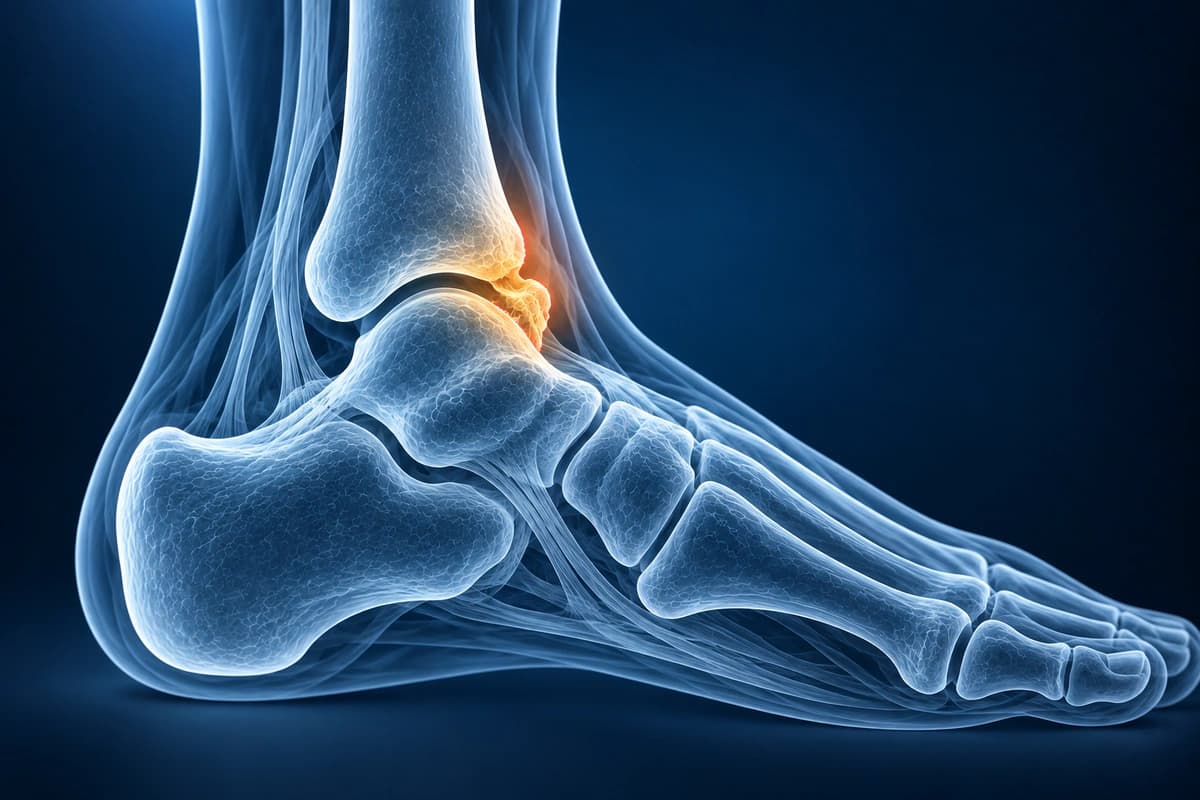
Ankle impingement refers to painful compression of bone or soft tissue during ankle motion, most commonly at the anterior or posterior tibiotalar joint. Anterior impingement typically results from repetitive dorsiflexion trauma causing osteophyte formation on the distal tibia and talar neck, often seen in athletes such as footballers and dancers. Posterior impingement involves compression of structures behind the ankle during plantarflexion, frequently associated with an os trigonum or elongated lateral tubercle of the talus. Soft tissue impingement can also occur from synovial hypertrophy, meniscoid lesions, or scarred capsular tissue. MRI is the preferred modality for evaluating both osseous and soft tissue causes of impingement. AI can help explain these findings in plain language, but clinical decisions require an ankle specialist to review the scan and examine you.
What We Look For on Imaging
- Anterior tibial and talar neck osteophytes causing mechanical abutment
- Synovial thickening and hypertrophic soft tissue in the anterior recess
- Os trigonum or Stieda process with surrounding bone marrow edema posteriorly
- Posterolateral soft tissue edema and flexor hallucis longus tenosynovitis
- Meniscoid lesion or anterolateral soft tissue mass in the lateral gutter
- Tibiotalar cartilage changes and subchondral cystic degeneration at contact zones
Frequently Asked Questions
How does MRI differentiate anterior bony from soft-tissue ankle impingement?
Anterior bony impingement is identified on sagittal and coronal CT or MRI by osteophytes on the anterior tibial plafond and dorsal talar neck, with contact visible in dorsiflexion. On MRI, sagittal PD-FS images demonstrate osteophyte size, subchondral marrow edema at the contact zone, and cartilage loss at the tibiotalar joint. Soft-tissue anterolateral impingement shows a hypertrophic synovial band or scar tissue in the anterolateral gutter — appearing as intermediate T1 and low-to-intermediate T2 signal — without osseous prominences. Ankle MRI with the foot in mild dorsiflexion can accentuate contact and confirm diagnosis.
What is os trigonum syndrome and how is it diagnosed on imaging?
Os trigonum is an accessory ossicle at the posterior talar process that impinges between the posterior tibial plafond and calcaneus during forced plantarflexion. On lateral radiograph, a distinct round ossicle separate from the posterior talar process is diagnostic. MRI adds critical information: STIR hyperintensity within the os trigonum and surrounding soft tissues indicates active impingement, and fluid within the synchondrosis between the os and posterior talar process on T2-FS confirms pathological stress. FHL tenosynovitis frequently co-exists, identified as sheath fluid and intratendinous signal on axial sequences.
Can AI-assisted MRI analysis predict which impingement patients respond to injection versus surgery?
AI can help explain report terms about bone spurs, thickened joint lining, and cartilage damage. It cannot predict whether an injection or surgery will help you. An ankle specialist must assess the scan together with your symptoms and examination before discussing treatment.
Sources
- Robinson P, White LM. Soft-tissue and osseous impingement syndromes of the ankle: role of imaging in diagnosis and management. Radiographics. 2002;22(6):1457-1469.
- Hedrick MR, McBryde AM. Posterior ankle impingement. Foot Ankle Int. 1994;15(1):2-8.
- Tol JL, Slim E, van Soest AJ, van Dijk CN. The relationship of the kicking action in soccer and anterior ankle impingement syndrome. A biomechanical analysis. Am J Sports Med. 2002;30(1):45-50.
- van Dijk CN, Tol JL, Verheyen CC. A prospective study of prognostic factors concerning the outcome of arthroscopic surgery for anterior ankle impingement. Am J Sports Med. 1997;25(6):737-745.
Get an educational explanation of imaging terms related to Ankle Impingement
Upload your MRI or X-ray DICOM files for private, AI-powered analysis. 4 models analyze independently — raw scan files stay in your browser.
Upload & AnalyzeMedical Disclaimer: This page is for informational and educational purposes only. It does not constitute medical advice, diagnosis, or treatment. AI-generated analysis may contain errors. Always consult a qualified healthcare professional for medical decisions. Full Disclaimer